


Specifications and Applications
Fuji Photo Film Co., Ltd. 26-30, Nishiazabu 2-Chome, Minato-ku, Tokyo 106-8620, Japan, Tel: +81-3-3406-2201, Fax: +81-3-3406-2158 • http://lifescience.fujifilm.com • E-mail: sginfo@tokyo.fujifilm.co.jp
Fujifilm Medical Systems U.S.A., Inc. 419 West Avenue, Stamford, CT 06902, U.S.A., Tel: +1-203-324-2000 ext. 6112 (1-800-431-1850 ext. 6112 in the U.S.), Fax: +1-203-351-4713 • http://www.fujimed.com • E-mail: SSG@fujimed.com
Fuji Photo Film (Europe) GmbH, Heesenstr. 31, 40549 Düsseldorf, Germany, Tel: +49-211-5089-174, Fax: +49-211-5089-139 • http://www.fujifilm.de • E-mail: mkaling@fujifilmeurope.de
Specifications and system configuration subject to change for improvement without notice. All other product names mentioned herein are the trademarks of their respective owners.
Ref. No. BB-204E (04,09)
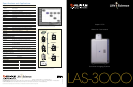
LAS-3OOO
S u p e r C C D
R e m o t e C o n t r o l l e d
S c i e n c e I m a g i n g S y s t e m
Image Capturing Unit (Camera Head and Controller)
CCD chip Fujifilm Super CCD Area Type chip
Number of pixels 3.2M pixels
Pixel size 10.75 x 10.75µm
Cooling Two-stage thermoelectric module with air circulation
Cooling temperature -30°C (when room is below 28°C)
Focusing Power focusing; remote and preset control
Exposure time 1/100 second to 30 hrs (2 hrs to 30 hrs to be set manually)
Dynamic range Four orders of magnitude
Gradation 16 bits (65,536)
Image size 12.6MB Max.; 49.2KB Min.
Maximum sample size 25 x 25cm (wide angle lens); 14 x 21cm (Fujinon VRF43LMD lens)
Binning 1 x 2, 2 x 4, 4 x 8, and 8 x 16 pixels
Interface USB1.1
Software
Image capture Fujifilm Image Reader (Mac
™
and Windows
®
)
Image analysis Fujifilm Image Gauge (Mac
™
); Fujifilm MultiGauge (Windows
®
)
Dimensions and Weights
Camera head 180 (W) x 170 (H) x 250mm (D) 3.4kg
Dark Box IV 510 (W) x 730 (H) x 480mm (D) 49.0kg
Analyzing Unit
Operating system Windows
®
XP or Mac
™
OS 9 & X
Applicable Reagents and/or Samples
Chemiluminescence CDP-Star
®
, ECL
™
, ECL Plus
™
, SuperSignal, ImmunoStar, CSPD
®
Fluorescence EtBr (W/UV light), SYBR
®
Green I & II, SYPRO
®
Orange,
GFP, DY-458XL (blue LED)
Pro-Q
™
Diamond, Cy
™
3, RFP, DY-520XL (green LED), Alexa Fluor 633,
Cy
™
5, DY-647 (red LED)
Chemifluorescence *AttoPhos
™
Digitization CBB-stained gel, Silver stained gel (Transilluminator)
NBT/BCIP-stained membrane. MW marker (Epi-illuminator)
Intelligent Dark Box IV
EPI-illuminator for fluorescence blue LED (470nm), green LED (520nm), red LED (630nm)
EPI-illuminator for digitization White-light source
Transilluminator - for digitization of
stained gels and autoradiographic films White LED
UV transilluminator** UV-light source (312nm)
Filter turret Five positions
Printers
Pictrography 3500
Lens Lens
High-sensitivity lens (FUJINON VRF43LMD) Wide-view lens
F-number 0.85 F-number 2.0
Focal length 43mm Focal length 24mm
Focus Remote power focusing Focus Manual
Mount Bayonet Mount Adapter to Nikon F mount
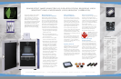
Analysis
Set a tray
Remote controlled
focusing
Exposure
Sample is placed on a tray
Chemiluminescence
Fluorescence
Chemifluorescence
Digitization
Chemiluminescence Mode
Fluorescence
Chemifluorescence Mode
Digitize Mode
Sample
Filter
blue LED
green LED
red LED
UV trans-
illuminator
White light
epi-illuminator
White trans-
illuminator
The image analysis process captures images in three modes:
chemiluminescence, fluorescence/chemifluorescence and digitization.
The binning mode of the LAS-3000 allows researchers to select from four
binning settings to enhance both imaging sensitivity and image resolution.
*No license is granted for use of AttoPhos™ to detect nucleic acid on a nylon membrane.
**No license is granted for pre-labeling gel electrophoresis method with the UV transilluminator.