


C H E M I L U M I N E S C E N C E • F L U O R E S C E N C E / C H E M I F L U O R E S C E N C E • D I G I T I Z A T I O N
The LAS-3000 imaging system combines
Fujifilm’s high-sensitivity Super CCD
camera technology with the added ver-
satility of white, blue, green and red EPI
illuminators. The Super CCD imaging
chip, binning mode and specially designed
camera lens allow researchers to capture
faint-light luminescent images with
unprecedented sensitivity and resolution.
Multicolor illuminator options enlarge
application area in fluorescent imaging.
Super CCD - By rotating pixels 45 degrees
to form an interwoven layout, the Super
CCD’s pixel pitch in the horizontal and
vertical directions is narrower than in the
diagonal direction, achieving higher hori-
zontal and vertical resolution.
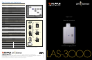
NP Tray
Fujifilm Super CCD Area
Type imaging chip
FUJINON lens VRF43LMD and
five-position filter turret
The five filter options available
for the LAS-3000.
More sensitivity for
chemiluminescent detection
Western blotting, Southern blotting and
Northern blotting detection by chemilumi-
nescence is a widely accepted method. The
use of a cooled CCD camera system enables
the generation of a digital image and quanti-
tative analysis of the image’s signal strength.
Several state-of-the-art technologies were
incorporated to make the LAS-3000 system
as sensitive as the conventional film method:
• The newly designed F0.85 high-sensitiv-
ity Fujinon lens gathers as much light as
possible onto the CCD.
• The CCD is a 3.2M-pixel Super CCD
having large octagonal-shaped CCD pixels
10.75 x 10.75µm in size.
• The CCD is cooled to -30°C by peltier
when the environment is below 28°C.
• Pixel binning in the LAS-3000 electroni-
cally increases the pixel area to increase
sensitivity. The binning mode includes
four binning levels: 1 x 2 (Standard),
2 x 4 (High), 4 x 8 (Super) and 8 x 16
(Ultra). In the Ultra mode, the exposure
time is nearly 60 times faster than
Standard. An image smoothing function
increases the pixels to make the image
size the same as the Standard.
• Long exposure times are required to
capture images of extremely low-light
samples. The low-noise design of the
LAS-3000 system includes default settings
for a two-hour exposure and a 15-hour
exposure in addition to manual settings
for exposures as long as 30 hours.
• A non-parallax (NP) tray is used to elimi-
nate the parallax effect when imaging
chemiluminescence in the 96-well plate.
Up to two plates can be placed.
LED units
More versatility for
fluorescent detection
More and more fluorescent dyes are intro-
duced into the biochemistry and molecular
biology fields for staining proteins and
nucleic acids in gels, membranes and well
plates. Expression analysis using green fluo-
rescent protein (GFP) and red fluorescent
protein (RFP) can be detected by blue exci-
tation or green excitation respectively.
The LAS-3000 includes multiple LED
options for various fluorescence applications,
including blue LED (470nm), green LED
(520nm) and red LED (630nm) for epi-illu-
mination and a UV (312nm) light source for
DIA-illumination.
The five-position filter turret holds 77mm
filters, such as Y515 (general purpose for
blue LED excitation), 510DF10 (GFP, etc.),
575DF20 (RFP, Cy
™
3, Pro-Q
™
Diamond,
etc.), R670 (Cy
™
5, ALEXA Fluor
®
633, etc.)
and 605DF40 (EtBr, etc.).
Digitizing functions
Capturing images as we see them is called
digitizing. For CBB-stained gel or silver-
stained gel, use of the white LED DIA-
illuminator at iris f2.8 without any filter is
recommended. For digitizing the molecular
weight standard on blotted membrane, white
light epi-illuminator, which is located in the
center part of the blue, green and red LED
epi-illuminator unit, is used.
Easy-to-use operation
Samples are placed on the appropriate tray
and placed in the intelligent dark box.
After closing the door, all the functions are
controlled remotely from the Image Reader
software. The software automatically rec-
ognizes the type of illuminator installed or
changed during operation.
Users are able to control the LAS-3000
with the Image Reader software in either
the user-friendly Lite mode or the more
advanced Pro mode. The modes are
selected with the click of a mouse.
Quantitative image
analysis function
The 16-bit signal accumulated on each
CCD pixel is processed through an A/D
converter to form a digital image. The
image data obtained at the closed shutter
condition is the dark frame. The image
data of an even, flat sample is the flat
frame. To analyze the image data, the dark
frame and flat frame data are subtracted
from the original image. This correc-
tion function is included in the Image
Reader software. MultiGauge software
for Windows
®
and ImageGauge software
for Mac
™
are the standard image analysis
software included in the ScienceLab
software provided by Fujifilm.
Applications
Fluorescence/Chemifluorescence
Blue: SYBR
®
Green I & II SYBR
®
Gold
SYPRO
®
Ruby SYPRO
®
Orange
SYPRO
®
Tangerine FITC
FAM™
EGFP
ECFP AttoPhos™
DY-458XL
Green: SYPRO
®
Red Cy
™3
TAMRA™ ROX™
HEX™ Alexa Fluor
®
532
Alexa Fluor
®
546 Deep Purple
Pro-Q™ Diamond Rhodamine Red™
BODIPY
®
576/589 NED™
R-phycoerythrin RFP
HNPP DY-520XL
DY-547
Red: Alexa Fluor
®
633 Alexa Fluor
®
635
Alexa Fluor
®
647 Cy
™5
BODIPY
®
650/665 DiD
TOTO
®
-3 DDAO phosphate
DY-647
UV: Ethidium Bromide SYPRO
®
Rose
White: Silver stained CBB
NBT / BCIP X-ray film
Chemiluminecence
ECL™
ECL Plus
™
ECL Advance
™ Lumi-Light Plus
Super Signal
®
CDP-Star™
CSPD
®
Renaissance™
Bright-Star™
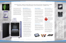
Luminescent image analyzer (LAS) for applications requiring high
sensitivity and a wide range of fluorescent capabilities
GFP expression in tobacco leaf
Detection of EPO as doping substance in sports
Isoelectric profiles of natural EPO found in normal
human urine (B) and the bands found in urine after
injections of epoetin or darbepoetin.
Data courtesy of Dr. Françoise Lasne.
Laboratoire National de Dépistage du Dopage,
Châtenay-Malabry, France