


28
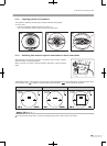
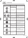
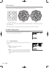
Examples of retinal photograph Possible cause Remedies
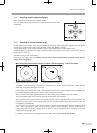
Dark shadow or bright reflection
appears at the top or bottom of the
image.
The instrument is positioned
too high to the patient’s eye.
Perform all alignment steps to obtain a clear
retinal image with consistent brightness. De-
pending on the patient’s eye fixation condi-
tion, you may obtain a good image by locating
working dots away from the dot position aid.
The instrument is positioned
too low to the patient’s eye.
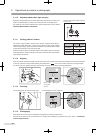
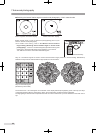
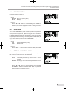
Dark shadow or bright reflection
appears at the left or right of the
image.
The instrument is positioned
too right or left to the patient’s
eye.
Perform all alignment steps to obtain a clear
retinal image with consistent brightness. De-
pending on the patient’s eye fixation condi-
tion, you may obtain a good image by locating
working dots away from the dot position aid.
Diopter compensation knob is
not in click.
Check that diopter compensation knob is in
the correct position by slowly moving the knob
to see if it cricks.
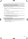
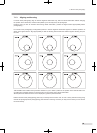
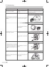
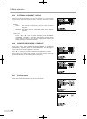
Peripheral area become whitened.
The instrument is positioned
too close to the patient’s eye.
The instrument is positioned
too far from the patient’s eye.
Locate the digital camera to the position
where working dots become smallest. When
flares still persist, pull the digital camera a
little or move the optical component to the
patient. (Working dots may be fades a little.)
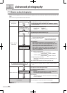
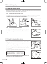
The image is too dark.
The pupil diameter is small.
Turn exposure compensation knob to +1 or
+2 position. If the patient’s pupil diameter
equal to or smaller than that shown by the
working dots, press SP mode button and
photograph in SP mode.
The image is out of focus or
blurred.
The patient has developed a
cataract.
Avoiding the area of white turbidity during
alignment may allow you to obtain a good im-
age.
The corneal surface is dry. Ask the patient to blink before imaging.
The image become whitened lo-
cally.
The objective lens is dirty.
Clean the objective lens.
Refer to
“10. Maintenance and inspection”
for details of the objective lens cleaning.
The lower area become whitened.
The eyelid or eyelashes intrude
the imaging area.
Ask the patient to open his/her eyes wide so
that the eyelid or eyelashes do not intrude
the area of the pupil, or you or your assistant
help the patient keep the eye wide open.
8
Troubleshooting